Elbow Anatomy
Online Biology Dictionary|
|
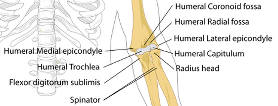
The seven basic elements of elbow anatomy are the bones, the articular cartilages, the ligaments, tendons, muscles, blood vessels, and nerves.
The elbow joint (Latin: articulatio cubiti) connects the upper arm bone (the humerus) with the two bones of the forearm, the ulna and the radius. The radius is smaller than the ulna and connects to the wrist on the same side as the thumb.
Basic Elbow Anatomy
The elbow is basically a hinge joint, or ginglymus, but it also can twist as the radius turns against the humerus, which occurs with rotation of the hand. The head of the radius is cup-shaped so that it can turn against the capitellum, the ridge that constitutes humerus's contact point with the radius. The trochlea of the humerus is received into the semilunar notch of the ulna. The surfaces where these three bones come into contact are coated with articular cartilage, a smooth substance that allows the bones slide painlessly against each other. Articular cartilage is white, rubbery. shiny smooth.
Ligaments
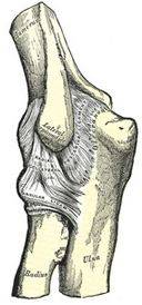
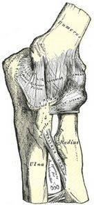
The bones of the elbow are held together by ligaments. The main ligaments of the elbow (illustrated below) are:
- The radial collateral ligament, or lateral collateral ligament, which lies on the lateral surface of the joint and connects humerus with the ulna and with the annular ligament. This ligament is generally triangular in shape (see illustration).
- The ulnar collateral ligament, or Medial collateral ligament, which lies on the medial surface of the joint and connects the inner end of the ulna with the inner end of the humerus.
- The annular ligament, which wraps around the radial head (the word annular means "ring-shaped"). The annular ligament cinches the radius to the ulna without interfering with the rotation of the radial head.
- The posterior ligament, which lies on the posterior surface of the joint and also attaches the end of the ulna with the humerus
- The anterior ligament, which lies on the anterior surface of the joint
A thin layer of tissue known as the joint capsule fills the spaces between the ligaments and forms a waterproof chamber. This chamber is filled with synovial fluid, a lubricating liquid that eases movement of the joint.
Click on these illustrations to enlarge them:
|
|
| |
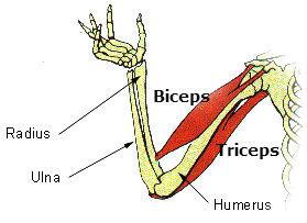
| The biceps muscle inserts on the radius just distal to the elbow, while the triceps inserts on the anterior surface the ulnar head. |
- The biceps tendon, which attaches the biceps muscle to the forearm, inserts in (is attached to) the upper radius near the elbow. The biceps muscle flexes the arm, as in when you do a pull-up.
- The triceps tendon, which attaches the triceps muscle to the forearm, inserts at the upper end (head) of the ulna near the elbow. The triceps muscle straightens the arm, as in when you do a push-up.
- Most of the tendons of the muscles that straighten the fingers and wrist insert on lateral epicondyle of the humerus, the prominence (or bump) on the outer surface of humerus immediately adjacent to the elbow.
- The muscles that flex the fingers and wrist come together in a tendon that inserts on on medial epicondyle of the humerus, the prominence that forms the bump on the inner surface of the elbow.
Nerves
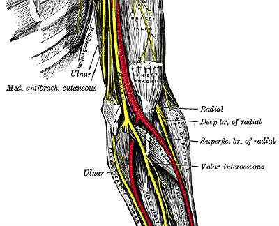
| Elbow anatomy: Major nerves |
|
- The radial, which lies on the same side of the forearm as the thumb, has both a deep and superficial branch.
- The volar interosseous, which lies between the two bones of the forearm (the ulna and the radius), arises as a branch of the median.
- The ulnar nerve, which runs within the medial portion of the forearm, adjacent to the ulna.
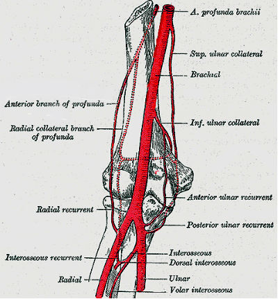
| Elbow anatomy: Arteries |
|
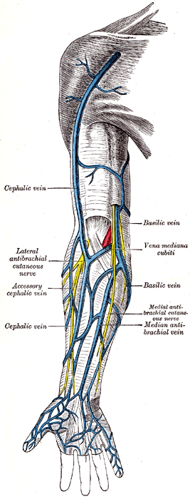
| Elbow anatomy: Veins |
|
Most shared on Macroevolution.net:
Human Origins: Are we hybrids?
On the Origins of New Forms of Life
Mammalian Hybrids
Cat-rabbit Hybrids: Fact or fiction?
Famous Biologists
Dog-cow Hybrids
Georges Cuvier: A Biography
Prothero: A Rebuttal
Branches of Biology
Dog-fox Hybrids
