Exocytosis
Online Biology Dictionary
|
|

|
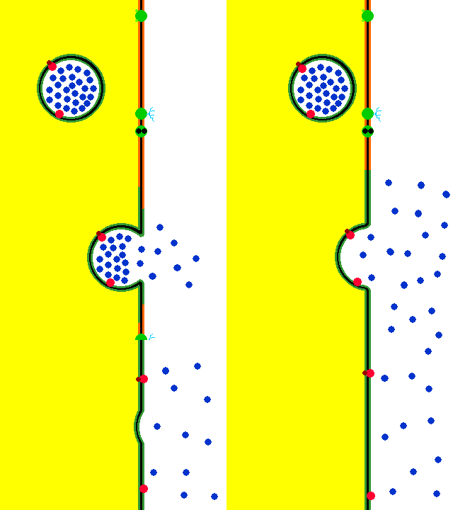
| Exocytosis animation. Used with permission. Courtesy Chris Patton, Stanford Sea Urchin Embryology Lab |
Exocytosis, sometimes called "reverse pinocytosis," is the process by which macromolecules are expelled from a cell into the extracellular environment ("secreted"). In the animation at right, the yellow region is the interior of the cell and the white area represents the exterior. During the course of this expulsion, the macromolecules, which are shown in blue in the animation, are first sequestered within a vesicle (represented at right, by the green circle). The vesicle then moves to, and fuses with, the plasma membrane. The contents of the vesicle are the molecules to be secreted. Membrane proteins and lipids produced within the cell are also sent by this process to become part the plasma membrane.
In the animation, the surface proteins of the plasma membrane are shown in red, those of the vesicle membrane, in green. Note that when the vesicle fuses with the plasma membrane, the membrane's proteins are displaced by the surface proteins of the vesicle and the molecules in the vesicle are expelled. So, really, the red and green regions are composed of the same surface proteins, the different colors are only for the purpose of clarifying how the process occurs.
When this process occurs on an ongoing basis, it is termed "constitutive." If, however, an external stimulus is required to stimulate its occurrence, it is called "regulated."
In nerves, synaptic transmission occurs when the pre-synaptic neuron secretes neurotransmitter molecules, which then travel across the synaptic cleft to receptors on the postsynaptic neuron.
Most shared on Macroevolution.net:
Human Origins: Are we hybrids?
On the Origins of New Forms of Life
Mammalian Hybrids
Cat-rabbit Hybrids: Fact or fiction?
Famous Biologists
Dog-cow Hybrids
Georges Cuvier: A Biography
Prothero: A Rebuttal
Branches of Biology
Dog-fox Hybrids