Kinetochores
Chromosome connection points
Online Biology Dictionary
|
|
EUGENE M. MCCARTHY, PHD

|
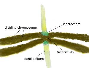
Kinetochores (/kə-NET-ə-korz/). The kinetochore is the structure where the spindle apparatus attaches to a chromatid during cell division (mitosis and meiosis) in eukaryotes. It is usually located in or near the centromere.
| Etymology: The prefix kineto- is from the Greek word kinesis, meaning movement. The suffix -chore is from the Greek word choros, meaning place. |
In mammals, when the spindle connects, 15-35 pole-to-chromosome microtubules ("kinetochore microtubules") attach to each single kinetochore. But the number is smaller in many other organisms. In yeast only a single microtubule connects. Each normal chromatid has one and only one kinetochore.
Like other biological structures, kinetochores are composed of protein. They not only play a role in connecting the chromosomes to the microtubules, but also in propelling them along them (by means of motor proteins, such as dynein and kinesin, that they contain).
A single kinetochore is composed of two regions: (1) the inner kinetochore, which is permanent and directly associated with centromere DNA; and (2) the outer kinetochore, which interacts with the microtubules and only assembles during cell division.
An extremely cool video about chromosomes and kinetochores: