Microtubules
Online Biology Dictionary
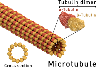
Microtubules (/MIKE-rō-TOOB-yoolz/) are hollow strands of tubulin, which are polymerised from α- and β-tubulin dimers (see figure at right below). They are present in all eukaryotic cells, where they are found in undulipodia and cilia. They also make up part of the cytoskeleton that maintains the structure of cells. In addition, they form the spindle apparatus during cell division, the focus of discussion here.
 A microtubule and the alpha- and beta-tubulin heterodimer it's constructed from. There are 13 protofilaments surrounding the hollow center. Image: Thomas Splettstoesser
During mitosis and meiosis there is a disassembly of the cell's cytoskeleton and the spindle apparatus forms, which is basically an outgrowth of large numbers of microtubule (MT) filaments from the centrosomes, which form the two centers of the apparatus. For this reason, the centrosomes are referred to as the microtubule organizing centers (MTOCs). They are duplicated during G2 of interphase, so there are two centromeres present in each dividing cell as mitosis begins. Within the MTOC is a third, distinct type of tubulin, designated as γ-tubulin.
Three types of microtubules
As the spindle apparatus forms, a microtubule can take on three possible roles. It can become a kinetochore microtubule, a polar microtubule, or an astral microtuble. Strictly speaking, only the first two of these three types (kinetochore and polar) make up the spindle apparatus, but all three play an essential role in moving the chromosomes.
A kinetochore microtubule, as its name suggests, is the type of microtubule that attaches to the kinetochores of the condensed chromosomes (this is the type that draws the chromosomes toward the centrosomes at the poles of the dividing cell).

|
|
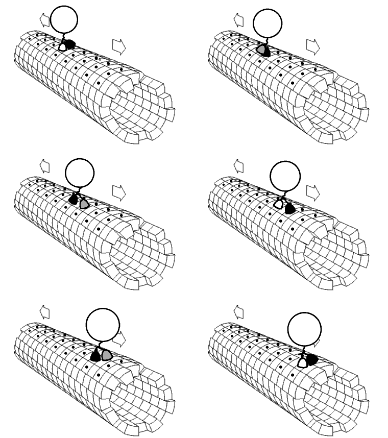
The motor protein kinesin walking a microtubule. Source: Wikimedia |
A polar microtubule does not attach to a chromosome. Instead, they emanate from the two centrosomes and overlap with each other in the center of the cell and, by sliding against each other, force the centrosomes toward the poles of the cell.
Astral microtubules extend outward from the centrosomes forming an aster (Greek for star) of radiating filaments which reach to the cell membrane. The wall-attached filaments pull the centrosomes toward one of the poles.
Motor proteins
Motor proteins are molecular motors, that is, they are tiny, molecular-scale machines that bring about various types of movement in living cells. A motor protein consumes energy in the form of ATP and converts it into motion or mechanical work. Kinesin and dynein are two types of motor proteins found in eukaryotic cells. They attach themselves to MT filaments and walk along them (see animation). MT-based molecular motors play a role in mitosis. Kinesins, in particular, are important for regulating the length of the spindle apparatus. They are involved in sliding the MT filaments apart within the spindle during metaphase, and in shortening the filaments during anaphase by depolymerizing their ends at the centrosomes, which draws the chromosomes to the poles. Different families of kinesin proteins have different actions. For example, the kinesin-5 family slides the microtubules apart within the spindle, whereas the kinesin-13 family depolymerizes microtubules at the centrosomes.